The kidneys are innervated by nerves relaying information from the central nervous system to the kidney (efferent) and nerves relaying the state of kidney function to the central nervous system (afferent). Although studies in preclinical models as well in clinical studies in humans undergoing catheter-based renal nerve ablation suggest both may contribute to hypertension, very little is known regarding the underlying mechanisms.
Anatomy of afferent renal nerves (collaboration with Dr. Lucy Vulchanova)
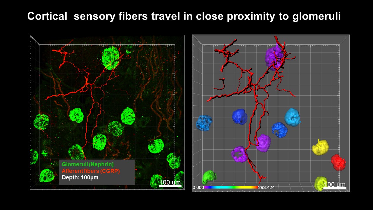
The Osborn lab has partnered with Dr. Lucy Vulchanova in the Department of Neuroscience at the University of Minnesota to combine anatomical studies of renal nerves with physiological investigations. This partnership is currently funded by two grants from the National Institutes of Health. Roman Tyshynsky, a PhD student in the Graduate Program for Neuroscience, has been conducting anatomical studies as part of this thesis project. Although it is commonly believed that renal sensory nerves innervate the renal pelvic wall primarily, current studies using a variety of state-of-the-art-methods, are revealing that renal sensory nerves are also in the renal cortex and are in close approximation to renal glomeruli. The figure below illustrates a 100µm-thick section of immunohistochemically labeled renal cortex (left), with glomeruli shown in green (Nephrin) and afferent nerve fibers in magenta (CGRP). 3D mathematical models of these afferent fibers and the glomeruli (right) allow for the quantification of the relationship between the afferent fibers and glomeruli. For example, here the glomeruli are colored to represent their proximity (µm) to the bundle of afferent fibers. We hypothesize these nerves provide important sensory information to the central nervous system related to glomerular function. We are currently testing this hypothesis using a variety of approaches.
_______________________________________________________________________________________________________________________________________________________
Targeted ablation of afferent renal nerves in a hypertension model of renal inflammation
We are currently investigating how renal nerves interact with inflammation in the kidney. These studies have been led by Dr. Chris Banek during his time as a post-doctoral fellow in the lab. We hypothesize that under some conditions inflammatory cytokines within the kidney activate renal afferent nerves resulting in chronic activation of the sympathetic nervous system and hypertension. Direct recording of afferent renal nerve activity (ARNA) in a preclinical model of hypertension with established renal inflammation has clearly demonstrated increased nerve discharge. In addition to increased ARNA in this model of hypertension, targeted ablation of renal afferent nerves, afferent renal denervation (ARDNx) attenuated the development of hypertension by 50% and to the same degree as total (afferent + efferent) renal denervation (TRDNx).
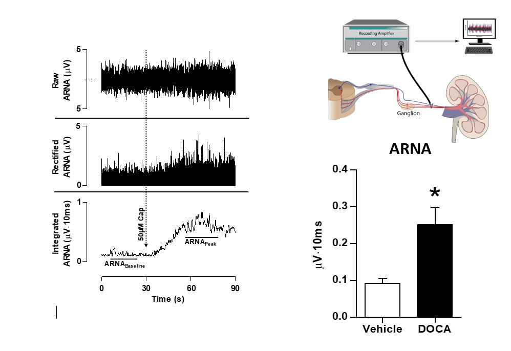
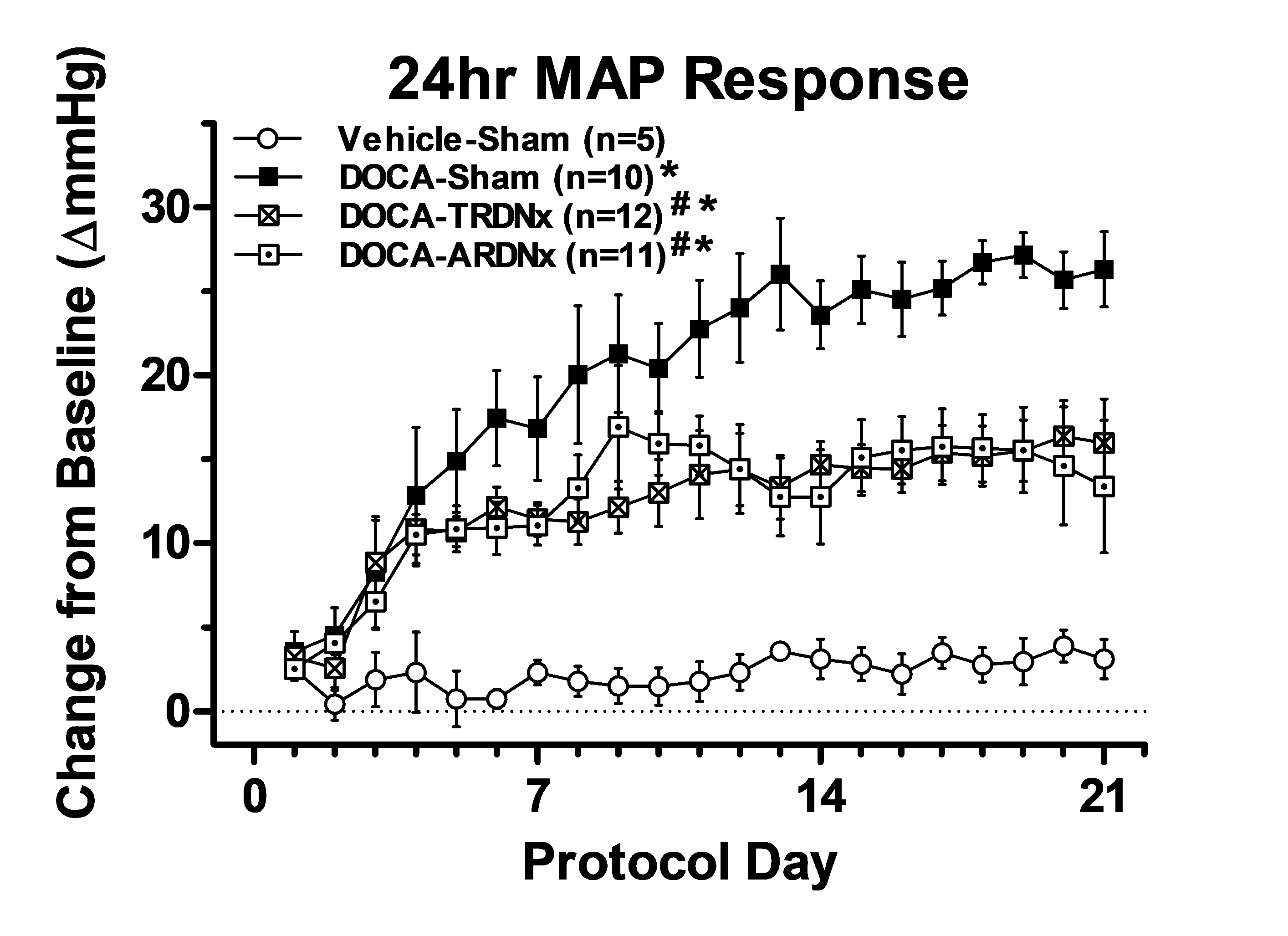
From: Resting afferent renal nerve discharge and renal inflammation: Elucidating the role of afferent and efferent renal nerves in DOCA-salt hypertension. Banek et al, Hypertension 68: 1415-1423, 2016.
_______________________________________________________________________________________________________________________________________________________
Anatomical and physiological investigations of the relationship between renal inflammation and afferent renal nerves (collaboration with Dr. Lucy Vulchanova)
The anatomical relationship between immune cells, the cytokines they secrete and afferent renal nerves is largely unknown. We are investigating the anatomical relationshipbetween immune cells and renal afferent nerves. Also, studies are currently underway to establish whether the receptors that mediate the cellular actions of inflammatory cytokines are present on sensory nerve terminals of renal afferent nerves.
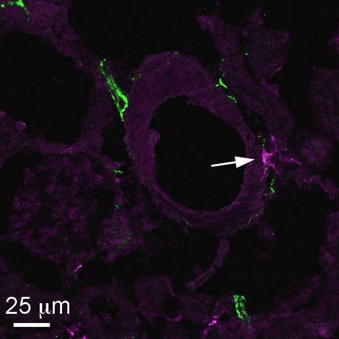
Arrow indicates association of a CGRP-positive (afferent) nerve fiber (green) with an Iba1-labeled
macrophage (purple) at the surface of a renal blood vessel in a DOCA-salt hypertensive rat.